We can develop and provide equipment and imaging software that feed back customer feedback, so we will propose the most efficient X-ray CT equipment that matches the sample.
Equipped with abundant shooting modes such as half scan, full scan, normal scan, offset scan, intermittent scan, continuous scan, and multi-stage scan, it realizes high-speed shooting of 20 seconds * at the fastest 30 fps and high-speed reconstruction of the fastest 4 seconds.
* May vary depending on the combination of devices.
From sample set to 3 cross-section CT image display, one-touch simple shooting operation, sample centering on a personal computer can be performed, and difficult shift value adjustment can be automated with the automatic shift value calculation function
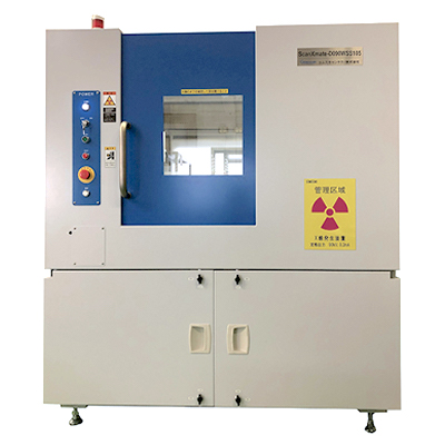
Our X-ray CT equipment responds to the problems of various customers such as food processing companies, pharmaceutical companies, machine manufacturers, battery manufacturers, research institutes, etc.
We shoot a wide variety of samples under optimal conditions and provide images that satisfy our customers.
If you have any questions, please feel free to contact us.

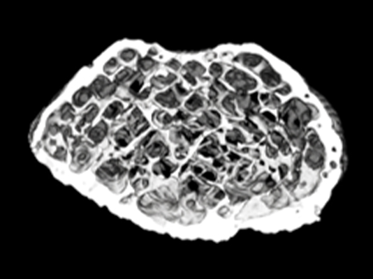
I want to check if there is any foreign matter in the package
(Food processing company)

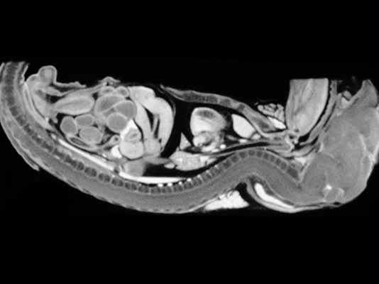
I want to check the distribution structure of chemicals inside the capsule
(Pharmaceutical company)

I want to shoot a mouse with an X-ray CT scan method that reduces the burden on the living body.
(Research institute)
An X-ray examination (simple X-ray examination) image is a two-dimensional image that is projected onto a flat surface regardless of the thickness of the body. Therefore, the information in the thickness direction of the body is missing.
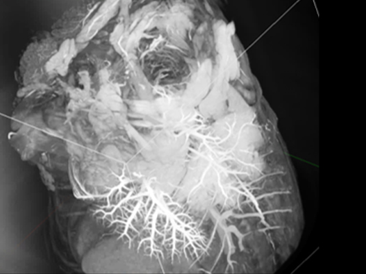
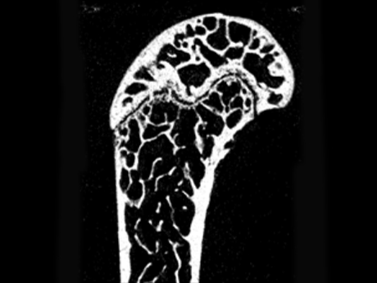
The X-ray CT image is a fluoroscopic image that is irradiated with X-rays from all directions of the inspection target and is generated as a three-dimensional image by reconstruction calculation. Since you can get location information in all directions, you can use it for the following purposes.
●You can accurately visualize and observe the inside of an unknown object without destroying the sample.
●Foreign matter inside the object and defects such as voids and cracks can be extracted without destroying the sample.
In addition, these areas, volumes, and positions can be measured accurately.
●3D dimensional measurement can be performed easily and accurately.
It can be compared with the design value and the wear and deterioration status can be quantified.
●You can create visually easy-to-understand images and videos from the data of the object to be shot.
●Can be used for 3D printers as STL data.
●t can be used for product quality judgment by sampling inspection and in-line inspection.
[Differences in shooting methods]
| Shooting method | Merit | |
|---|---|---|
| Continuous scan | A shooting method that captures images by shooting at the same timing as the number of views during continuous rotation of the stage. | High-speed shooting is possible. |
| Intermittent scan | Number of views A shooting method that repeats stage rotation ⇒ stage stop ⇒ image acquisition ⇒ stage rotation ⇒ stage stop ⇒ image acquisition for each pitch. | Suitable for high quality shooting. |
[Shooting time (example)]
| Binning | Frame rate | Number of views | Shooting time |
|---|---|---|---|
| 1×1 | 30fps | 1200 | 50 seconds |
| 1×1 | 15fps | 1200 | 90 seconds |
| 2×2 | 60fps | 600 | 25 seconds |
| 2×2 | 30fps | 600 | 30 seconds |
| explanation | |
|---|---|
| Manual shift value calculation | The center of the X-ray detector is set as the default value of the center of rotation of the sample, and the CT image is created while shifting the center of rotation to the left or right (+,-) in 1-pixel units. This is a method of visually determining the sharpest outline of an image and using that position as the shift value. |
Automatic shift value calculation |
The range of 50 pixels left and right (+,-) from the center of the X-ray detector is defined as the shift value judgment area, and the shift value is driven in the direction where the mean value of the histogram of the CT image is maximized, and the shift value is automatically set in 0.1 pixel units. It is a method to confirm. |
The NVIDIA Quadro RTX 4000 8GB high-speed graphics board delivers the industry’s fastest fast reconstruction operations.
| Image size | Reconstruction operation time |
|---|---|
| 512×512×512 600 views full area |
< 4 seconds |
| 992×992×992 1200 views full area |
< 26 seconds |
Shooting conditions can be selected from professional operation mode and easy operation mode.
Professional operation modes that can change the shooting conditions for each shot sample are X-ray tube voltage, X-ray tube current, binning, frame rate, number of views, number of integrations, sample physical position (X-axis, Y-axis, Z-axis, Φ-axis). ) Etc. can be freely set by the operator.
Since the imaging conditions after setting can be saved and the conditions can be read out and set arbitrarily, it is more convenient when taking the same sample or similar sample after a period of time.
After setting the shooting conditions, with one click of the button, calibration data collection ⇒ shooting ⇒ automatic shift value calculation ⇒ reconstruction calculation ⇒ 3 section display is fully automatic.
When the same sample is taken continuously, the operation is left to the device to some extent, which reduces the burden on the operator.
Various application software can be bundled.
Please contact us if you are considering application software other than the above.